Projekt
Untersuchungen auf Basis µ- und nano-CT
Untersuchungen auf Basis von µ- und nano CT für Artenidentifizierung und Strukturanalytik
Seit 2017 beschäftigt man sich im Thünen-Institut für Holzforschung im Arbeitsbereich 1 (Qualität von Holz und Holzprodukten) mit der Frage, inwieweit sich die µ- und nano-CT Technologie im Feld der Artenidentifizierung und Strukturanalytik einsetzen lässt. Dabei geht es insbesondere um die Identifizierung von Hölzern, bei denen prozessbedingt keine DNA mehr erhalten ist oder die chemotaxonomische Signatur verändert wurde wie beispielsweise Holzkohle oder verschiedene Werkstoffe aus Holz. Hintergrund ist, dass hier beispielsweise keine genetischen Untersuchungen mehr möglich sind und somit letztlich nur die anatomische Identifizierung relevant ist. Darüber hinaus wird die CT-Technologie bereits erfolgreich im Feld der Strukturanalytik eingesetzt. Beispielsweise konnte die Ausprägung einer besonderen Holstruktur, der “Riegelung” im Bergahorn und deren Holzbildungsdynamik im Xylem und Phloem, untersucht und Charakterisiert werden
Hintergrund und Zielsetzung
Mikro-CT ist eine hochauflösende, zerstörungsfreie Bildgebungsmethode, die es seit den 1990ern gibt und die heute in Form verschiedenster Instrumente angeboten wird. Zur Identifizierung von Hölzern, bzw. von organischen Mikrostrukturen allgemein, bietet sich insbesondere die hochauflösende sub-µ- und nano-CT Technologie an. Dort werden millimetergroße Proben auf dünne Szintillationsschirme projiziert und von einem optischen Mikroskop abfotografiert. Daraus entstehen hochauflösende (volumetrische) 3D-Bilder der Holz-Mikrostruktur, die es erlauben, sich virtuell durch die Struktur zu bewegen, ohne den Umweg über die aufwendige Probenpräparation zu gehen. In Kooperation mit dem Fraunhofer-Entwicklungszentrum Röntgentechnik EZRT und dem Thünen-Institut für Holzforschung ist es nun erstmalig gelungen, volumetrische Darstellungen auf Basis von µ- und nano-CT Daten mit künstlicher Intelligenz (maschinelles Lernen) zu verknüpfen und im Feld der Holzartenidentifizierung anzuwenden.
Zielgruppe
Wissenschaft, Umweltschutz, Politik und Handel
Vorgehensweise
Im Folgenden werden eine Reihe aktueller Studien in der Originalform (Abstracts) aus den wissenschaftlichen Publikationen dargestellt:
Study I: Volumetric imaging by micro computed tomography: a suitable tool for wood identification of charcoal (Haag et al 2023).
The present study focuses on the application of state-of-the-art µCT, by using a sub-micrometer CT scanner as a tool for wood identification. Charcoal was chosen as a subject for this case study. The reason for choosing charcoal is based on economic as well as technical issues. Parallel to conventional wood anatomy, various promising approaches to identification are currently being developed worldwide in order to simplify the identification of processed wood. However, due to the carbonization process, such approaches are not applicable to charcoal. In view of the rapid development of µCT technology, it was decided to examine the extent to which wood anatomical studies can be supported and improved by modern µCT technology. About 17% of the annually harvested wood worldwide is converted to charcoal (FAO 2017), and the charcoal trade is one of the least controlled/monitored segments of the European timber market. Although charcoal has a significant market share of wood-based products, it is still not yet covered by any trade regulation, e.g. the European Timber Regulations (EUTR), (EU) No995/2010. For the present study, different wood types and the anatomical fine structural features were measured and displayed at different magnifications to visualize the performance of state-of-the-art µCT standards. Three different charcoal assortments were examined and the results checked against the given declarations of contents. The aim of this work is to evaluate the potential of the µCT technique in the field of wood identification and to assess its use for the regulatory control of charcoal and other wood products in the international timber trade. The results are encouraging and lead to the conclusion that the application of the µCT technique in the field of wood identification can be classified as very promising for the future.
The presented method is generally applicable for wood identification and should be in the focus of wood anatomy, especially for products where chemotaxonomic or DNA based methods are unsuitable. In some cases, wood anatomy can be reliably performed at the species level. A considerable advantage results from the fact that for a selected fragment the method is non-destructive and the fragment is completely preserved. Subsequently, the volumetric image can be digitally screened and examined in detail, simplifying the search for relevant diagnostic features. The complex production of thin microtome sections is not necessary. Due to the high-resolution potential, the method can be used successfully as a tool for any kind of wood anatomical investigation. Thanks to the very small fragment size, the study is transferable to the investigation of any kind of wood fragments, e.g., for chips of materials like particle board or MDF. A possible scenario would be, for example, that modern CT machines based on artificial intelligence (deep learning) would be able to detect individual fragments, digitally separate them and identify the used wood. However, further research in this field is still necessary. The authors of the present study have already started working on a research project to establish the method. This will not only help pave the way for future applied wood identification, but also contribute to international cooperation and collaboration in this field. Finally, based on the results of this study, the application of-state-of-the-art µCT by using a detector-based sub-micrometer CT scanner as a tool for wood identification as presented here can be classified a suitable technique for wood identification.
Study II: Applications in the scope of anatomical wood identification using sub-µCt based volumetric images (Haag et al 2023).
Current developments and advances in the field of digital image processing/analysis and neural network programming (machine learning) open up new research perspectives and applications for wood anatomy and wood quality research. In the last decades, traditional wood anatomy has, against all expectations, experienced a renaissance and gained increasing importance in the field of wood identification. This is highly significant for the strict implementation of the laws governing the timber trade (CITES, EUTR, FLEGT, etc.)5, especially when other methods are basically not applicable due to process related changes in the wood (chemical modification, destruction of DNA). Various teams worldwide are currently engaged in taking traditional wood anatomy into the digital age in new fields of application. The main focus here is the use of neural networks. The first machine vision-based wood identification systems were primarily concerned with the macroscopic analysis of transverse sections of commercial timbers. In the course of the further development of these techniques, the resolution of the examined areas became higher and made observations at the microscopic level possible, yet so far limited to cross-sections4, 8, 3, 5, 7. In a recent study, the authors address the use of high-resolution sub-µCt based volumetric images. The potential of µ-CT technology is currently being explored for a variety of applications1,2 and the extent to which three-dimensional representations can be used in the field of artificial intelligence is being investigated.
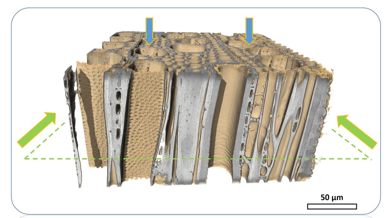
Study III: Wavy grain in sycamore maple (Acer pseudoplatanus)– a structural analysis of xylem and phloem
Wavy grain (fiddle back) in trees has attracted the interest of both science and industry due to its aesthetic and economic importance. As the underlying causes of this particular structural feature in trees remain uncertain, this study investigates the possibility of non-invasively detecting wavy grain patterns within tree bark tissue of sycamore maple (Acer pseudoplatanus). Wavy grain, characterized by alternating light and dark stripes on the wood surface, is highly valued in the production of musical instruments. This research explores the potential of employing transmission light microscopy and microcomputer tomography (μCT) to examine the internal structure of phloem tissue to inspect whether wavy grain structure found in the xylem continues in the bark.
The investigation of phloem tissue proved challenging due to its irregular cellular arrangement. Nonetheless, slight phloem fiber deviations were observed in individual trees. However no definite connection was found between the periodic oscillations of the xylem fiber tissue and the structure of the phloem in a given area. Variability in wavy grain expression among the xylem of the trees posed an additional challenge, with amplitudes and wavelengths differing across specimens. The application of μCT offered advantages over traditional sample preparation, enabling the study of the anatomical structure across larger areas and multiple depths without altering the specimen.
This study sheds light on the potential of employing advanced imaging techniques to detect wavy grain patterns, providing insights that could benefit industries reliant on high-quality wood, such as musical instrument crafting and forestry.
Study IV: Anatomical characterization of NTFPs (Non-Timber Forest Products/Shells and Nuts) for species identification (please also see NTFPs-Project)
The background to the project is the anatomical characterization of organic structures in the sense of an identification key. The studies on charcoal and briquettes carried out regularly since 2016 at the Thünen Center of Competence for Wood Origin show that a significant increase in substitution products of charcoal can be observed. While fragments of organic material were initially detected that could not be reliably assigned until then, parallel studies were carried out on important NTFPs in order to characterize them anatomically and differentiate them reliably from charred wood tissue. The problem here is that the relevant materials, such as coconut and walnut shells or olive pits, consist largely of sclereid cells and are extremely hard. Preparation of thin sections with a slide microtome, as is usually done for wood anatomical studies, is largely not applicable here. For this reason, a study is currently being carried out in cooperation with the Fraunhofer Development Center X-ray Technology EZRT to characterize and describe the cellular structure of important NTFPs, which is additionally based on alternative modern examination methods. The material under investigation is analyzed microscopically using diverse methods. please also visit: (hier wird in Kürze ein Link eingefügt, der zum “NTFP-Projekt” auf der Projektseite führt).
Literature:
Haag V, Kirsch S, Koch G, Zemke V, Richter H-G, Kaschuro S. 2018. Non-destructive investigation of historical instruments based on 3D-reflected-light microscopy and high resolution µ-X-ray CT. In: Wooden musical instruments - different forms of knowledge, book of end of WoodMusICK, COST Action FP1302. pp 143-156
Haag V, Dremel K, Koch G, Zabler S. 2023. Applications in the scope of anatomical wood identification using sub-µCt based volumetric images. In: IUFRO Div 5 Conference : The forest treasure chest: delivering outcomes for everyone ; 4-8 June 2023, Cairns, Australia.
Haag V, Dremel K, Zabler S. 2023. Volumetric imaging by micro computed tomography: a suitable tool for wood identification of charcoal. IAWA J 44(2):210-224, DOI:10.1163/22941932-bja10106
Lewandrowski T, Dremel K, Haag. 2024. Wavy grain in sycamore maple (Acer pseudoplatanus)– a structural analysis of xylem and phloem. IAWA J. (under review/march 2024).
Links und Downloads
Links:
https://www.researchgate.net/publication/370943755_Forensic_wood_identification_in_the_digital_age
Thünen-Ansprechperson

Thünen-Beteiligte
Zeitraum
Daueraufgabe 9.2017 - 9.2029
Weitere Projektdaten
Projektstatus:
läuft


